Breadcrumb
- News and Events
- News
- Content
- Interview with Dr. Barry Bedell
null Interview with Dr. Barry Bedell
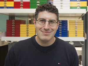
FACE TO FACE with Dr. Barry Bedell: A multimodal vision for the SAIL Platform - Scientist, Metabolic Disorders and Complications (MeDiC) Program, Research Institute of the McGill University Health Centre (RI-MUHC) - Director, Small Animal Imaging Labs (SAIL) Platform, RI-MUHC - Associate Professor, Department of Neurology and Neurosurgery, McGill University |  |
Below is an interview with a scientist who revels in constantly pushing himself to stay on top of what’s happening in his field, which is no small feat in neuroscience. Your current research focuses on Alzheimer’s and Parkinson’s diseases, which are thought to be caused in part by proteins that do not fold into their correct shape. How does non-invasive animal imaging help you investigate these protein-misfolding diseases? In adults with Alzheimer’s disease, a protein called beta-amyloid, in addition to another protein called tau, become misfolded in the brain. These misfolded proteins are suspected to cause neurons within the brain to eventually die. As the disease progresses, misfolded proteins spread to multiple regions of the brain. The same goes for Parkinson’s disease, where the misfolding of the alpha-synuclein protein is associated with the disease’s relentless progression to more and more severe problems with movement and dementia. The toxic effects of misfolded tau (called tauopathy) or alpha-synuclein (synucleinopathy) can be visualized in mouse models for these diseases. With multimodal imaging we can understand the disease process better, and we can also create new tools, called biomarkers, that help us to assess the status of the disease and see how well it is responding to treatment. What kind of multimodality assessments do you perform on mouse models? For Alzheimer’s and Parkinson’s models, we focus on a number of things that are known to change with the onset of disease in humans. These include the obvious ones, such as gait and brain structure, but also the metabolism of glucose, the brain’s energy source. Interestingly, these diseases often affect the ability of patients to sleep and smell, and lead to cognitive decline. With multimodality imaging, we can see how the parts of the brain that contribute to sleep, olfaction, and cognition are compromised in these disease models. What are the advantages of using non-invasive brain imaging in research? Non-invasive imaging allows us to study the same animal at multiple points in time. This longitudinal approach reduces the number of research animals used. It also improves our understanding of how pathology changes over a lifetime. For example, we can witness how parts of the brain degenerate and how glucose levels are altered. The major advantage of working with animal models is that we can, ultimately, establish a direct relationship between data gathered from a living animal, vs. data gathered from isolated organs and cells. This correlation is difficult or often impossible to do in humans. As such, we have a better understanding of what the images we obtain actually reflect at the level of cells and molecules. Now fully operational, the RI-MUHC’s SAIL Platform is unique in the world in the breadth of imaging modalities it can offer. What makes it so special? SAIL is one of very few facilities with such a broad spectrum of sophisticated imaging modalities and functional assessment tools, and it attracts scientists from many disciplines, including cardiology, infectious diseases, orthopedics, oncology, neurology and respiratory diseases. We’re a one-stop shop that offers the latest in MRI, CT, PET, SPECT and optical imaging. What sets the SAIL Platform apart is that it can complement these imaging studies with instruments to measure specific features of each animal’s gait while walking, or its quantity and quality of sleep. Your interest in brain research was sparked during your doctoral work, when you figured out ways to quantify the effects of multiple sclerosis using MRI scanners. Why the brain, and how did it lead you to oversee non-invasive imaging at the RI-MUHC? In my opinion, the brain is one of the most interesting organs to study using imaging because you can apply all available modalities, from brain macro- and micro-structure to cerebral blood flow to neurochemistry. After wrapping up my PhD at The University of Texas Health Science Center and MD Anderson Cancer Center in Houston, I began medical school at McGill, where I eventually piloted animal imaging in human MRI scanners because, at that time, there was no animal MRI scanner in Quebec. Following my residency in Anatomical Pathology, I founded the first small animal imaging lab at the Montreal Neurological Institute and Hospital. Now we have a truly unique, world-class research platform at the RI-MUHC. What motivates you outside of the realm of research? I thrive on understanding what’s beyond the periphery by staying attuned to the latest technologies on the market, especially rapid advances in computing and state-of-the-art innovations in other fields. How would you sum up your career to date? I’m doing exactly what I want to do and what I’ve always enjoyed. I don’t think that anyone can ask for more than that. — September 2017 |