Breadcrumb
- News and Events
- News
- Content
- Discovery of new genes for molar pregnancies sheds light on their increased incidence in women aged 35 and over
null Discovery of new genes for molar pregnancies sheds light on their increased incidence in women aged 35 and over
Major advances made at the RI-MUHC could help improve counselling for individuals suffering from infertility, premature ovarian insufficiency, recurrent miscarriages and androgenetic hydatidiform mole.
Montreal, November 18, 2024 — A molar pregnancy, also known as a hydatidiform mole, is an abnormal human pregnancy with no embryo and an overgrowth of the cells that form the placenta. The common form of molar pregnancies affects one in every 600 pregnancies in Quebec. Half of these moles are androgenetic, that is, they contain only the father’s chromosomes with no chromosomes from the mother, and their frequency increases 10 times with advanced maternal age. Because of the hyperproliferation of their cells, androgenetic moles may become malignant and lead to a placental cancer in up to 15 per cent of cases.
Scientists at the Research Institute of the McGill University Health Centre (RI-MUHC) recently discovered six new genes – FOXL2, MAJIN, KASH5, SYCP2, HFM1 and MEIOB – that cause recurrent androgenetic moles, recurrent miscarriages and infertility when mutated on both alleles (copies of the same gene) in the patients. Five of these genes are essential for Meiosis I, the process of cell division necessary for the production of sperm and eggs in humans. Previous studies have linked defects in some of the six genes to premature ovarian failure, a well-known cause of female infertility. In addition, five of these genes have been linked to male infertility.

These findings, just published in The Journal of Clinical Investigation, will improve the molecular diagnosis of recurrent molar pregnancies, premature ovarian failure and infertile women and men.
“Our findings suggest that recurrent androgenetic moles are a sign of ovarian ageing. They will change current clinical practice by introducing the evaluation of ovarian reserve for patients with recurrent moles,” says Rima Slim, PhD, corresponding and co-senior author of the study, Senior Scientist in the Child Health and Human Development Program (CHHD) at the RI-MUHC and Professor in the Department of Human Genetics at McGill University.
The six new genes add to four other genes that are also responsible for recurrent molar pregnancies and that were previously discovered by the same team (NLRP7, discovered in 2006, and MEI1, TOP6BL and REC114, discovered in 2018).
A broad international investigation
In collaboration with the team led by Jacek Majewski, PhD, Investigator at the RI-MUHC and Professor of Human Genetics at McGill, the researchers performed exome sequencing on 75 unrelated patients referred by physicians from around the world. These patients had at least two hydatidiform moles and did not have mutations in the previously described genes associated with the condition.
The researchers then checked whether the patients who were negative for biallelic mutations (on both alleles of a gene), had only one defective allele in genes with roles in Meiosis I and ovarian functions. They added 240 patients with other forms of reproductive failure - referred primarily from the MUHC Repeated Pregnancy Loss clinic, founded by Dr. William Buckett, and the Réseau des Maladies Trophoblastiques du Québec, founded by Dr. Philippe Sauthier. This second group of patients had either a molar pregnancy and at least one miscarriage, or at least two miscarriages without a molar pregnancy.
They found that 14 per cent to 28 per cent of these patients had one defective allele that appeared to be most frequent in patients with at least two molar pregnancies.
“Our data suggest that these monoallelic variants could be contributing, with other factors, to the genetic susceptibility of these patients for reproductive failure. Our study provides an explanation of the increased frequency of androgenetic moles with advanced maternal age,” explains Prof. Slim.
The authors of the study explain that “patients with monoallelic variants in these genes can conceive and have healthy children; however, they are at higher risk for infertility, premature ovarian insufficiency and reproductive loss than women from the general population.”
Modelling the genesis of moles
To better elucidate the mechanisms underlying this health and reproductive problem, the researchers modelled the disease in mice with deficiency in the HFM1 gene.
“We observed several defects that affect the meiotic progression, some of which were previously observed by our team in mice with deficiency in the Mei1 gene, another gene responsible for the causation of recurrent androgenetic moles,” explains Teruko Taketo, PhD, co-senior author of the study, Senior Scientist in the CHHD Program at the RI-MUHC and Professor in the Department of Surgery at McGill University. “In this study, using live-cell imaging, we were able to visualize and understand for the first time how the eggs from Hfm1 deficient mice lose all their chromosomes.”
The authors emphasize that the identification of the same mechanism in two mouse models supports its plausibility at the origin of androgenetic mole formation in humans.
“Androgenetic moles have been described in 1977. Today, we can better explain to the patients the formation of these aberrant conceptions and the genesis of androgenetic moles,” says Prof. Slim.
About the study
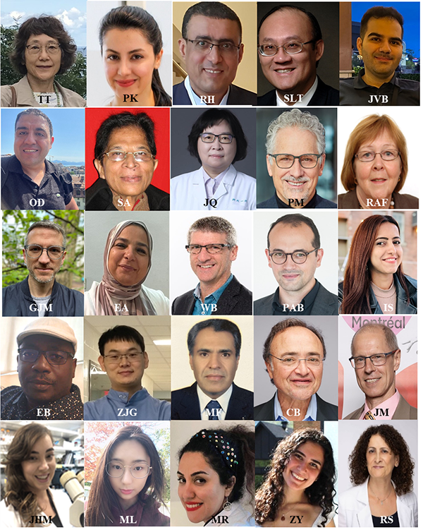
The study Defects in meiosis I contribute to the genesis of androgenetic hydatidiform moles was conducted by Maryam Rezaei, Manqi Liang, Zeynep Yalcin, Jacinta H. Martin, Parinaz Kazemi, Eric Bareke, Zhao-Jia Ge, Majid Fardaei, Claudio Benadiva, Reda Hemida, Adnan Hassan, Geoffrey J. Maher, Ebtesam Abdalla, William Buckett, Pierre-Adrien Bolze, Iqbaljit Sandhu, Onur Duman, Suraksha Agrawal, JianHua Qian, Jalal Vallian Broojeni, Lavi Bhati, Pierre Miron, Fabienne Allias, Amal Selim, Rosemary A. Fisher, Michael J. Seckl, Philippe Sauthier, Isabelle Touitou, Seang Lin Tan, Jacek Majewski, Teruko Taketo and Rima Slim.
DOI : https://doi.org/10.1172/JCI170669
This work was primarily supported by the Canadian Institutes of Health Research and also benefited from funding from Mitacs Accelerate in partnership with Originelle Inc., McGill’s Centre for Research in Reproduction and Development, the RI-MUHC Desjardins Studentship and the Faculty of Medicine and Health Sciences of McGill University.
The investigators would like to thank all the researchers and clinicians who contributed to the study, as well as the patients and families who participated.

About the Research Institute of the McGill University Health Centre
The Research Institute of the McGill University Health Centre (RI-MUHC) is a world-renowned biomedical and healthcare research centre. The Institute, which is affiliated with the Faculty of Medicine of McGill University, is the research arm of the McGill University Health Centre (MUHC)–an academic health centre located in Montreal, Canada, that has a mandate to focus on complex care within its community. The RI-MUHC supports over 720 researchers and close to 1,400 research trainees devoted to a broad spectrum of fundamental, clinical and health outcomes research at the Glen and the Montreal General Hospital sites of the MUHC. Its research facilities offer a dynamic multidisciplinary environment that fosters collaboration and leverages discovery aimed at improving the health of individual patients across their lifespan. The RI-MUHC is supported in part by the Fonds de recherche du Québec–Santé (FRQS). www.rimuhc.ca
Media contact
Fabienne Landry
Communications coordinator, Research, MUHC
FabiennePraesent id dolor porta, faucibus eros vel.landry@muhc.mcgill.ca
Related news